


Hip hip arthropathy is a degenerative dystrophic process that occurs in the joints of the femoral head and the pelvis and acetabulum. The disease is more typical in middle-aged and older adults, although it can also occur in younger people, including children. In most cases, its development is preceded by injury, as well as many pathologies of inflammatory and non-inflammatory nature, and pain and stiffness with movement become the main signs of the degenerative dystrophic process of the hip joint. During its development, the disease goes through several stages, if in the early stages it can be treated conservatively, then in the final stages, the treatment of hip arthropathy is only effective with surgery. Otherwise, the pathology will lead to severe disease or even complete immobilization.
What is Hip Arthropathy and Its Development Mechanism
Hip disease, also known as osteoarthritis and osteoarthropathy, is a complex disease of the hip joint (HJ) with progressive destruction of the cartilage. Over time, this causes the surfaces of adjacent bones to deform and form bone growths called osteophytes on them.
According to statistics, hip joint disease accounts for about 12% of all diseases of the musculoskeletal system. It is second only to knee arthropathy in terms of frequency, but has a much higher risk of disability.
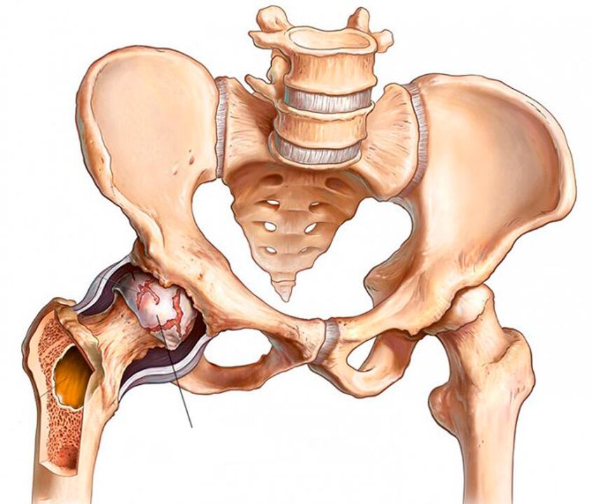
The two hip joints are the largest joints in the human body. Each of them consists of the femur and the acetabulum of the pelvis. The femoral head sits in a cup-shaped depression in the pelvis and can move freely in different directions. This joint structure allows flexion and extension, adduction and abduction, and rotation of the thigh.
To prevent discomfort from exercise, the surfaces of bones that touch each other are covered with an elastic layer called hyaline cartilage. It's him who makes the femoral head slide easily in the acetabulum. In addition, hyaline cartilage provides stability and cushioning of the hip joint during movement.
The entire joint is immersed in a shell called the joint capsule. It contains the synovial membrane of synthetic synovial fluid. It is she who lubricates the surface of the cartilage and ensures that water and nutrients flow into it, which is responsible for maintaining the normal structure of the cartilage tissue.
Above the joint capsule is a group of femoral and pelvic muscles that help the joint move. The hip joint is also surrounded by a set of ligaments that ensure the stability of its position within physiological boundaries.
Because the hip joint is subjected to heavy loads on a daily basis, it is prone to rapid wear and tear and injury. The risk of this change significantly increases the impact of many headwinds that are practically unavoidable in the modern world, but they are discussed below. This explains the high incidence of hip joint disease.
The production of synovial fluid is violated due to negative factors. Gradually, its quantity decreased and its qualitative composition changed: it became sticky, sticky and no longer able to adequately nourish the cartilage. This results in acute nutritional deficiencies and progressive dehydration of the hyaline cartilage. As a result of these changes, cartilage tissue loses strength and elasticity, and it flakes, ruptures, and loses volume. All of this prevents the smooth sliding of the femoral head in the pelvic acetabulum, leading to signs of hip arthropathy.
Gradually, the joint space narrows, increased friction occurs between the articular bone surfaces, and the pressure of the bone against the hyaline cartilage increases. This leads to greater injury and wear and tear, which cannot but affect the biomechanics of the hip joint and the health of the person.
Failure of the hip joint negatively affects not only the biomechanics of the lower extremity, but also the entire organ of movement. This often results in disability.
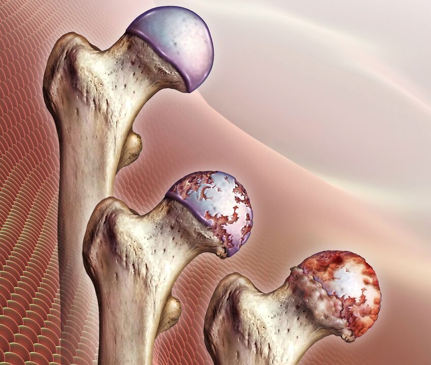
As the lesion progresses, the transparent layer gradually and completely disappears, resulting in the exposure of the bone surface and a dramatic increase in the load on the bone and joint. During movement, the femoral head is no longer covered by anything, but rubs directly against the pelvic-acetabular surface. In addition to severely limiting mobility and causing excruciating pain, the bones squeeze against each other while flattening out.
As the joint bone deforms, osteophytes (osteophytes) form on its surface. They can have sharp edges and seriously injure the surrounding muscles. This can cause severe pain in the groin, legs, and buttocks. As a result, the patient will subconsciously try to avoid the affected hip joint and avoid moving within it. Lack of adequate load on the muscles can cause them to gradually shrink, further exacerbating mobility problems. This leads to lameness.
Development reasons
Hip arthritis of the hip can be primary or secondary. In the first case, the reason for its development cannot be found, i. e. the disease develops on its own without an obvious cause. Secondary hip arthropathy is the result of many changes in the state of the musculoskeletal system or in lifestyle characteristics, in particular:
- Hip injuries, including fractures, dislocations, bruises, sprains or ruptures of surrounding ligaments, chronic microinjuries, etc. ;
- manual labor;
- a sedentary lifestyle;
- obesity;
- Chronic infectious processes in the body;
- Rheumatoid arthritis, gout, tendonitis, bursitis;
- endocrine disorders, metabolic and hormonal disorders, including diabetes;
- Congenital deformities of the hip (dislocation, dysplasia);
- aseptic necrosis of the femoral head;
- various spinal diseases;
- genetic susceptibility;
- Smoking addiction.
In the vast majority of cases, the development of hip osteoarthritis is due to unavoidable age-related changes, and the presence of the other factors mentioned above only increases the risk of its occurrence and increases the rate of progression.
symptoms and extent
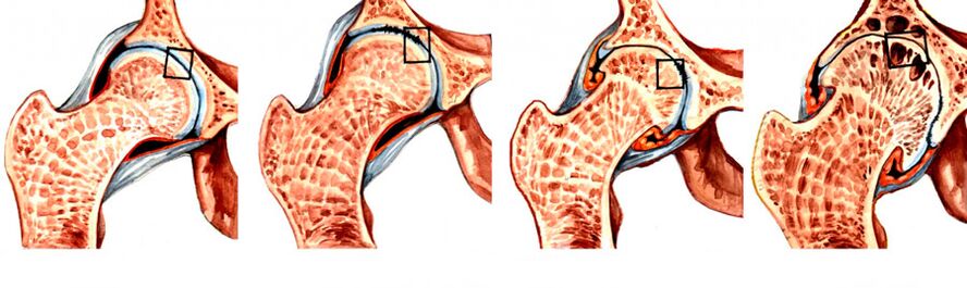
During hip arthropathy, 4 developmental degrees can be distinguished, 1 of which is the easiest. Initially, the disease may be asymptomatic or present with mild pain. They usually occur after intense physical exertion, long walks, or the end of a busy day. In the earliest stages of disease development, discomfort is often attributed to fatigue and is seen as the norm. Therefore, in rare cases, hip arthropathy is diagnosed in the first stages of development.
Visible signs of hip arthropathy begin to appear in the second stage of its progression, when the joint space is nearly halved and the femoral head is displaced and deformed. With the transition to stage 3, the pain becomes unbearable and disturbs a person even at night, and they tend to radiate to the buttocks, calves, groin, and buttocks. Since the joint space is virtually non-existent and multiple osteophytes have formed on the bone surface, independent movement is impossible in this case. Therefore, the patient is forced to use crutches or crutches.
Therefore, the main symptoms of hip osteoarthritis are:
- Limited mobility - Initially, patients may notice difficulty performing leg rotations, but over time morning stiffness and HJ swelling may also develop. Because of them, a person needs a few minutes to warm up, so to speak, and move around to regain a normal range of motion. Gradually, it becomes more and more difficult for the patient to perform leg exercises.
- Typical crunch - occurs when walking, and flexion or extension of the hip joint. This is the result of bone surfaces rubbing against each other, and is associated with a sharp or dull pain associated with hip arthropathy.
- Pain Syndrome - Pain initially after physical exertion and relieved by prolonged rest. Heavy lifting or hypothermia can trigger an exacerbation, as hip disease is often complicated by increased synovial inflammation. As the disease progresses, the pain becomes more frequent, lasts longer, and gets worse.
- Thigh muscle spasms - are the result of compressed nerves and weakened ligamentous tissue, so muscle spasms compensatory keep the femoral head in the acetabulum. Also, increased synovitis can cause muscle spasms.
- Lameness - Occurs in the final stages of disease development, as a deformation of the skeletal surface causes the appearance of flexor contractures. Therefore, one cannot fully straighten the leg and hold it in this position. Additionally, patients may limp involuntarily to transfer weight to healthy parts of the body, as this helps reduce the intensity of the pain.
- Shortening of the legs - Degree 3 hip arthropathy was observed. The leg may be shortened by 1 cm or more on the side of the affected hip due to narrowing of the joint space, decreased muscle tone, and flattening of the femoral head.
During the final stages of development, the femoral head fuses with the acetabulum, resulting in complete immobilization and disability of the leg.
At the same time, degenerative dystrophic changes can be observed in one hip or in both hips. Thus, characteristic symptoms will be observed on one or both sides, but in the latter case their severity may differ on the left and right sides.
diagnosis
Physicians can suspect hip arthropathy based on the patient's complaints, external examinations, and functional tests. During the visual inspection, be sure to measure the length of the legs. To do this, ask the patient to stand up and straighten their legs as much as possible. Measurements are taken between the anterior axis of the pelvis and any bony structure in the knee, ankle, or heel. However, the data obtained would not be informative if both hip joints were affected by hip arthropathy at the same time.
But because the typical symptoms of hip arthropathy may accompany many other inflammatory and non-inflammatory diseases, instrumental methods must be used to accurately diagnose the patient's pathology. It could be:
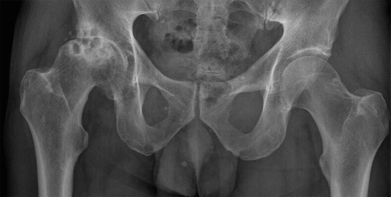
- CT or X-ray of the hip joint - the images show destructive changes, narrowing of the joint space, osteophyte formation and deformation of the bone surface;
- MRI is the most informative test and allows you to accurately assess cartilage structure, the nature of ligament changes, and the nature of blood circulation in the hip area.
Patients were also assigned laboratory tests to assess their general health and detect conditions that could lead to hip arthropathy. it:
- UAC and OAM;
- blood chemistry;
- Rheumatism check;
- Hip puncture and biochemical studies.
The task of diagnosis is to differentiate between hip and knee arthropathy (knee joint damage), as well as radicular syndrome that occurs with osteochondrosis, as well as disc herniation and herniation. In addition, the symptoms of hip arthropathy may resemble those of bursitis trochanus and the atypical course of ankylosing spondylitis, requiring a thorough examination to identify the real cause of pain and limited mobility.
Conservative treatment
Conservative treatment of hip joint disease is effective only in the initial stages of the disease. It is selected individually for each patient and may include a range of different approaches, each of which will complement the others. Therefore, as part of the treatment of hip osteoarthritis, patients may be prescribed:
- medical treatement;
- exercise therapy;
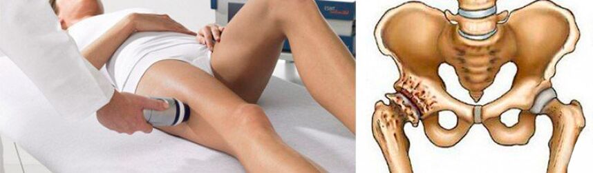
- physiotherapy;
- Plasma boost.
For conservative treatment to be effective, patients need to eliminate the influence of many factors that contribute to the development of hip arthropathy. If you are overweight, it is very important to reduce it as much as possible. This will reduce the load on the affected joints and the risk of progression of the degenerative dystrophic process.
You should also quit smoking, regulate physical activity, and avoid overloading, but don't sit all the time. To prevent further damage to the hip joint, special bandages and orthoses are recommended. They provide secure immobilization of the joint and support it during movement.
medical treatement
The nature of the drug treatment is strictly individualized. In most cases, patients are prescribed:
- NSAIDs - drugs that have both analgesic and anti-inflammatory properties (available in tablet, injection, and topical form);
- Corticosteroids - drugs with strong anti-inflammatory effects, prescribed if NSAIDs have no significant effect;
- chondroprotectant - helps activate the cartilage tissue regeneration process, but its effectiveness has not been proven;
- Muscle relaxants - drugs that reduce muscle tone and eliminate spasms, which are necessary when spasms of certain muscles or muscle groups against the background of severe pain;
- Preparations to improve blood circulation - most often in the form of an injectable solution, to help improve the nutrition of the tissues around the joints;
- B vitamins - shown to normalize the transmission of nerve impulses, which is especially important when nerves are compressed by deformed bone structures.
For acute pain that cannot be relieved by tablets, patients can be given intra- or peri-articular blocks. They are performed only by qualified health workers in medical facilities and involve the introduction of an anesthetic solution containing corticosteroids into the joint cavity or directly in the area around it.
exercise therapy
Therapeutic exercise is an effective way to deal with decreased muscle tone and limited mobility. Thanks to a properly chosen set of exercises, it is possible to increase the range of motion and reduce the severity of pain. If hip arthropathy is accompanied by pinching of nerve fibers, they also prevent muscle atrophy and help eliminate spasticity, which reflexively causes spasms of individual muscles.
Exercise therapy sessions improve blood circulation in the area of the degenerative dystrophic process. As a result, the nutritional quality of the diseased joint is increased and the regeneration process is accelerated.
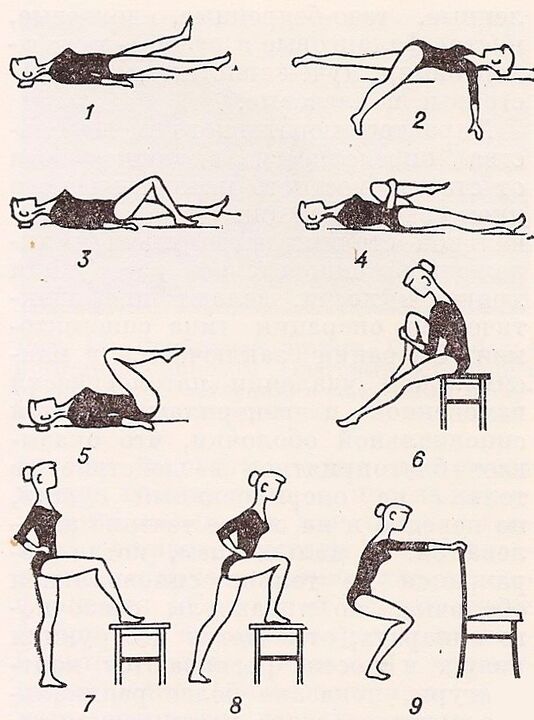
For each patient, a set of exercises should be developed individually by a specialist. At the same time, not only the degree of damage to the hip joint, but also the level of physical development of the patient should be considered.
physiotherapy
Physiotherapy procedures and massages are anti-inflammatory, analgesic, tonic and anti-edema. In addition, they help maintain normal leg muscle tone and prevent them from fatigue and atrophy.
For hip arthropathy, a course of 10-15 procedures is prescribed:
- ultrasound therapy;
- magnetic therapy;
- Laser Treatment;
- electrophoresis;
- Ultrasonic electrophoresis;
- UHF;
- Paraffin treatment.
In addition, many patients received mud therapy. Such surgery has a positive effect only in the first stage of the development of hip arthropathy or during the recovery period after surgical treatment. With the help of the therapeutic mud, it is possible to improve the quality of blood circulation and accelerate the recovery of the mobility of the affected joints.
Plasma Lift
Plasma lift, or PRP therapy, is a procedure in which platelet-rich plasma from the patient's own blood is introduced into the cavity of the hip joint. This allows you to activate the hyaline cartilage recovery process.
However, according to some scientists, such a process can lead to the formation of malignant tumors. This notion is based on the fact that plasma boosting promotes the formation of large numbers of stem cells, whose effects on the body have not been fully studied.
Surgical treatment of hip joint disease
Despite the obvious discomfort in the hip joint, many seek medical help too late, and by the time the joint disease reaches grade 3 or even 4 severity, function is irreversibly depleted.
With the development of pathology, surgery is a necessary measure. Only timely surgical intervention can help patients restore normal activities and save patients from extreme pain, that is, to achieve a significant improvement in the quality of human life. Without drugs, physical therapy procedures can restore severely damaged cartilage. At best, painful intra-articular injections and medications can reduce the pain. But this will be temporary, after which the pain will recur with the same or even greater intensity.
Indications for hip surgery are:
- joint space disappears;
- Persistent pain in the hip joint that cannot be relieved;
- severe mobility impairment;
- Hip fracture.
Depending on the severity of joint destruction and skeletal deformity, patients are offered various types of surgical treatment, namely:
- Arthrodesis;
- internal prosthesis;
- Osteotomy.
Arthrodesis
Arthrodesis is an affordable procedure that requires metal plates to firmly hold the joint bones. The result is complete immobilization of the joint. Therefore, with the help of arthrodesis, it is possible to correct only the support function of the leg, eliminate pain, but not have to talk about regaining mobility or a significant improvement in quality of life.
Today, arthrodesis is practically not used because it deprives a person of the opportunity to move fully.
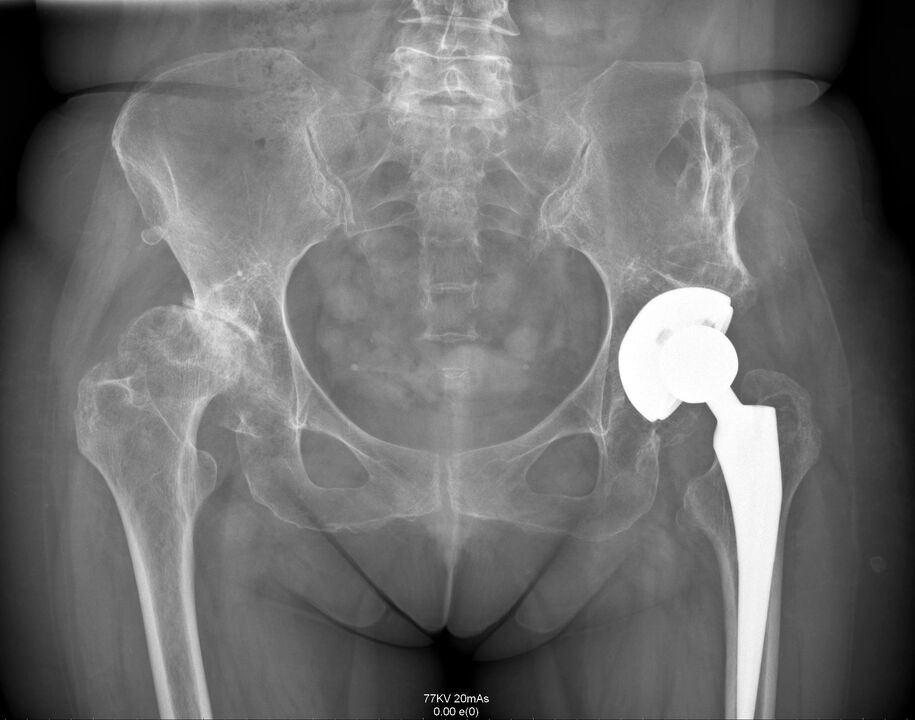
endoprosthesis
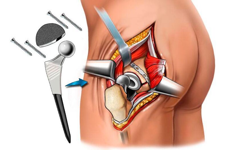
Arthroplasty is the only way to fundamentally address the hip joint problem and restore all its function and mobility. This is a high-tech solution to hip problems that will allow you to completely forget about it in 15-30 years, along with pain and mobility limitations. Thanks to the use of modern endoprostheses, it is possible to fully restore motor support functions and provide the patient with a normal life.
The surgery involves removing the femoral head and part of the neck. Surgical preparation of the acetabular bed was also performed, including removal of osteophytes, alignment of their surfaces, and excision of necrotic tissue. Endoprostheses can even be used to treat elderly hip patients.
The surgery is performed under general anesthesia and takes about an hour. Depending on the severity of the degenerative malnutrition process, it can be done using one of the following methods:
- Superficial - including grinding of the acetabulum and femoral head and further coating with smooth implants to replace the destroyed hyaline cartilage (this method is rarely used due to possible inflammation of the tissue surrounding the joint);
- Monopolar - the femoral head is removed and replaced with an endoprosthesis (used when cartilage remains on the surface of the acetabulum and only destroys the femoral head);
- Bipolar - similar to the previous technology, differing only in the design of the endoprosthesis used, which has a lower coefficient of friction and provides smoother motion on the joint bed;
- Total is the most effective and safest solution to hip problems in the hip, which involves complete removal of the femoral head and capture of part of the neck and acetabular fossa, with a full-fledged artificial joint.
Therefore, patients may be advised to install various types of endoprostheses. Most hip replacements are manufactured in the US and UK. For their manufacture, chemically and biologically inert metals are used: cobalt, chromium, titanium alloys. Ceramics are also commonly used. In most modern models, a polymer pad is additionally used, which provides the artificial TBS with natural shock absorption, stability and sliding properties.
When an endoprosthesis is performed, the success rate of the procedure is almost 100%.
After surgery, antibiotics are prescribed to prevent the development of infectious complications, and stitches are removed 10 days later. The postoperative scar size was about 8 cm, and the patient was discharged from the hospital. Rehabilitation after implantation is simple, but physical therapy, massage, and exercise therapy are still required.
osteotomy
An osteotomy is a surgical procedure that is a temporary measure before a total hip replacement with an artificial endoprosthesis. The essence of the surgery is to align the femoral axis due to an intentional fracture. The resulting fragments are placed in the most appropriate location, thereby slightly unloading the diseased joint. As a result, it is possible to temporarily reduce the severity of pain and improve mobility.
Therefore, hip joint disease is a rather scary disease that can completely deprive a person of the opportunity to move independently. It progresses for a long time, and its symptoms, especially early symptoms, are often regarded by patients as a normal state after physical exertion. But this is precisely the insidious nature of the disease, as it can only be managed non-surgically in the initial stages of its development. But if the degenerative dystrophic process has completely destroyed the hyaline cartilage and left the bone surface exposed or even flattened, only surgery can help the patient. Fortunately, the modern level of medicine and surgery in particular makes it possible to fully restore the hip joint to its normal state and function.